Hey Clubhouse! Sorry we've been out of the loop lately. We all just flew back in from San Diego, and boy are our arms tired.
One of our beloved colleagues from the community has recently declared to us his love of ultrasound guided nerve blocks--a skill that is established in the anesthesia world but is quickly gaining traction in our realm. Some programs are training their residents in this skill, and some aren't. But we've been inspired to talk about it! That said we will now be featuring a series of posts on this very subject. Our inaugural post today: femoral nerve blocks.
Let's say we do a little time warp, and we are unfortunately six months into the past, stuck in the dredges of frigid winter. EMS appears with a patient who just wiped out big time on the ice. He's gripping his thigh with his hands, and he is screaming. Maybe he's broken his hip? Maybe it's dislocated? Maybe he cracked the shaft of his femur? For this exercise, let's say that it doesn't matter.
What does matter is that it looks like this might need a little pushing and pulling, either to get it reduced or maybe just for re-positioning for imaging. Procedural sedation? I mean, we're always down for that, but is there a better way?
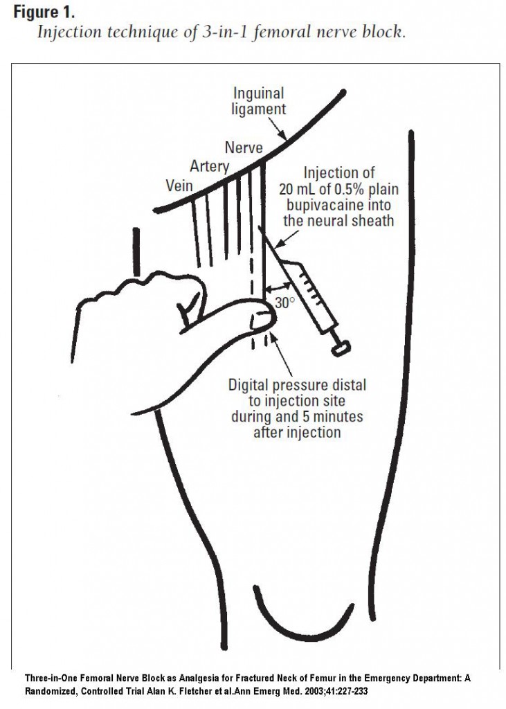
Turns out there is! Specifically we will discuss the femoral 3-in-1 block--so titled as it knocks out the lateral cutaneous, obturator, and femoral nerves. This will completely anesthetize the femoral shaft, with some coverage of the proximal end of the femur. This can be done blind, but adding U/S greatly increases the chance of success--the evidence that's out there has shown that using a probe gets you blockade faster and has a higher rate of complete blockade.
You'll need the following for your setup: an U/S machine with a linear probe, a 21 G spinal needle, a vial of 0.5% bupivacaine, sterile gloves, a sterile probe cover, and sterile gel.
Essentially your goal is to infiltrate the fascial sheath, allowing for the rapid distribution of anesthetic to the above mentioned nerves. And to properly do this procedure, you'll need a LOT of local anesthetic--our sources recommend up to 30 mL of bupivacaine.
When done blind, to confirm you'll either feel a gratifying "pop", your patient will get a paresthesia, or you will feel the pulsations of the femoral artery pushing your needle laterally. But we can do it better! For this jobby, prep and drape the area. Place your probe per convention in the transverse plane, starting at the inguinal ligament. Slide down until you can see the femoral vein and artery (about 2 cm caudad). You shouldn't miss these guys. Just lateral to them is the femoral nerve, which appears as a hyperechoic triangular structure.

Make a wheal for entry just lateral to the probe. Then go in with your spinal needle directed medially at a 30 degree angle. Get your tip as close as possible to the nerve, and then inject (well, after aspirating first, of course). You should see the local anesthetic spread the tissues in a cephalad direction. You'll want to hold pressure distally during all of this, and then 5 minutes after injection.
Here's a demonstration from the fine people at SonoSite:
Here's the usual video (with the usual hilarity) from Whit Fisher:
As always, these posts are for EDUCATIONAL PURPOSES ONLY. These nerve blocks have the potential to be huge for us as emergentologists, but don't take our word for it. Do your research, and always be safe.
jps/bmc
Sources:
Roberts and Hedges Clinical Procedures in Emergency Medicine. Saunders 2014, Chapter 31
Beaudoin, F. L., Haran, J. P., & Liebmann, O. (2013). A Comparison of Ultrasound-guided Three-in-one Femoral Nerve Block Versus Parenteral Opioids Alone for Analgesia in Emergency Department Patients With Hip Fractures: A Randomized Controlled Trial. Academic Emergency Medicine, 20(6), 584–591. doi:10.1111/acem.12154
Cristos, S., Chiampas, G, Offman, R, & Rifenburg, R. (2010). Ultrasound-Guided Three-In-One Nerve Block for Femur Fractures. Western Journal of Emergency Medicine. 11(4), 310-313
Cristos, S., Chiampas, G, Offman, R, & Rifenburg, R. (2010). Ultrasound-Guided Three-In-One Nerve Block for Femur Fractures. Western Journal of Emergency Medicine. 11(4), 310-313
Turner, A. L., Stevenson, M. D., & Cross, K. P. (2014). Impact of ultrasound-guided femoral nerve blocks in the pediatric emergency department. Pediatric Emergency Care, 30(4), 227–229.
Reid, N., Stella, J., Ryan, M., & Ragg, M. (2009). Use of ultrasound to facilitate accurate femoral nerve block in the emergency department. Emergency Medicine Australasia, 21(2), 124–130. doi:10.1111/j.1742-6723.2009.01163.xdoi:10.1097/PEC.0000000000000101
Fletcher, A. K., Rigby, A. S., & Heyes, F. L. P. (2003). Three-in-one femoral nerve block as analgesia for fractured neck of femur in the emergency department: A randomized, controlled trial. Annals of Emergency Medicine, 41(2), 227–233. doi:10.1067/mem.2003.51
SonoSite YouTube Page
SonoSite YouTube Page
Whit Fisher's YouTube Page

Hi,
ReplyDeleteYour blog is more informative. I appreciate your blog.
Keep sharing valuable information.
Regards,
Ultrasound guided Injection in Basildon
Hi,
ReplyDeleteThank you so much for your personal experience and to all the people who posted here. It has been a great help to me. This is definitely one of the best articles. I have read in this website! Thanks.
Regards,
Ultrasound guided Injection in Maidstone
Hey
ReplyDeleteAbsolutely brilliant.
really helpful.
please also read my blog:
Regards,
Ultrasound guided Injection in Bexleyheath